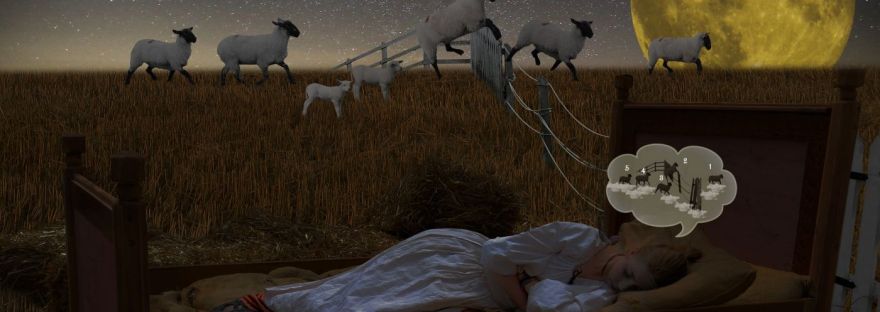
Psalm 3:5:“I lie down and sleep; I wake again, because the Lord sustains me.” (NIV)
A few other closely related passages include:
Psalm 4:8:“In peace I will lie down and sleep, for you alone, Lord, make me dwell in safety.” (NIV) / “I will both lie down in peace, and sleep; for You alone, O Lord, make me dwell in safety.” (NKJV)
Proverbs 3:24:“When you lie down, you will not be afraid; when you lie down, your sleep will be sweet.” (NIV)
The circadian clock is an internal, 24-hour biological timer located in the brain that regulates the body’s daily sleep-wake cycle and physiological processes. The circadian rhythm refers to the natural physical, mental, and behavioural changes that follow this daily cycle, responding primarily to light and darkness.
Key Components & Functions
The Master Clock: Located in the suprachiasmatic nucleus (SCN) within the brain’s hypothalamus, this master clock receives direct signals from the eyes regarding environmental light.
Hormone Regulation: Daylight signals the SCN to trigger the release of cortisol to promote alertness. Darkness prompts the production of melatonin, which signals the body to prepare for sleep.
Peripheral Clocks: Virtually every organ and tissue (such as the liver, heart, and gut) has its own cell-level clock synchronised by the master clock.
What It Influences
Biological Process
Peak Activity Time
Alertness & Core Body Temp
High during the day, drops significantly late at night
Digestion & Metabolism
Most efficient during typical daylight eating windows
Immune System Activity
Shifts repair and tissue restoration to overnight hours
Melatonin Production
Begins rising in early evening, peaking around 2–4 AM
Common Disruptions
Environmental Shift: Jet lag, night-shift work, and excessive exposure to blue light (screens) after sunset confuse the master clock.
Health Impact: Chronic misalignment between your schedule and your clock increases the risk of insomnia, metabolic disorders, depression, and cardiovascular issues.
Consistent sleep schedules, daily morning sunlight, and limiting bright screens before bed help keep the circadian rhythm aligned.
Comparing sleep and death is one of humanity’s oldest concepts, spanning neuroscience, literature, and theology. While sleep is an active biological repair process and death is a permanent cessation of life, they share deep physical, perceptual, and philosophical overlaps.
1. Physiological & Neurological Levels
Reduced Responsiveness: Both states involve a drastic drop in sensory awareness, muscle tone, and reaction to external stimuli.
Metabolic Slowdown: During deep, non-REM sleep, your core body temperature drops, heart rate slows, and breathing becomes long and shallow, mimicking the body’s physical presentation just before biological death.
Brain Activity Waves: Severe unconsciousness (such as a coma or general anaesthesia) sits on a continuum between sleep and brain death. In fact, general anaesthesia is often described as a medically induced, reversible coma rather than “true” sleep, serving as a physiological bridge between the two states.
2. Cognitive & Perceptual Levels
The “Lapse” of Consciousness: From the subjective perspective of the person, falling asleep involves a complete loss of ego and time perception. You do not experience the passage of hours while asleep; you simply wake up. Death, perceptually, is the permanent extension of that sudden non-experience.
Loss of Voluntary Control: In both states, the conscious self, surrenders control over the body, thoughts, and environment.
3. Linguistic & Cultural Levels
Euphemisms and Metaphors: Across languages, sleep is the primary euphemism for death. Phrases like “putting a pet to sleep,” “at rest,” or “rest in peace” rely entirely on this comparison to soften the reality of mortality.
Mythology: In Greek mythology, Hypnos (the god of sleep) and Thanatos (the god of death) were portrayed as twin brothers, highlighting how ancient cultures viewed the two conditions as mirror images.
Scriptural & Religious Texts: Many religious traditions treat sleep as a temporary form of death. In Christian scripture, death is frequently referred to as “falling asleep” (e.g., John 11:11, 1 Thessalonians 4:14), framing resurrection as an “awakening”.
The Key Difference
The fundamental divergence is reversibility and metabolic activity. Sleep is an essential, highly active neural state where the brain clears toxins, consolidates memories, and repairs tissue. Death is the irreversible breakdown of those metabolic and cellular processes. Sleep is a temporary withdrawal from consciousness to sustain life; death is the permanent end of it.
When a person goes to bed at night as a result of physical and or mental activities during the day, and falls asleep, subsequently after four to eight hours later, wakes up using an alarm clock, or not, is it possible to apply the scripture text, “I lie down and sleep; I wake again, because the Lord sustains me.”? Or is this simply a case of belief, in contrast to Knowledge of God or from God? How is it possible to know this? Since from the subjective perspective of the person, falling asleep involves a complete loss of ego and time perception. Interesting conundrums for everybody, including psychologist, psychiatrist, sleep therapist, and neurosciences.
John chapter 14 verse 6;Jesus answered, “I am the way and the truth and the life. Obviously this statement/text spoken by Jesus Christ is a metaphor, however, is there a literal meaning behind it? If so, let us examine the literal aspect. That is, instead of approaching ‘the life’ as a ‘who’, approaching it as a ‘what’.
Let us literarily examine what is life, how does it start? What is the technical process involved? Do we really know how life begins physically/technically? What do we know scientifically, and what do we not know (scientific ignorance)? What are the unproven theories we have in this field? And what are our best explanation of the quantum biochemical process of DNA replication.
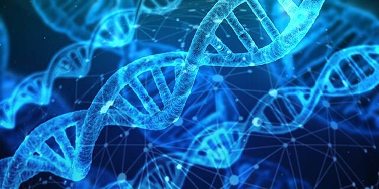
Deoxyribonucleic Acid (DNA) is the molecule that stores the genetic blueprint for all living organisms. It contains the instructions needed to build, operate, and reproduce every cell in the body. See picture below.
Structure of DNA
The Building Blocks — Nucleotides
DNA is a polymer made of repeating units called nucleotides, each consisting of three components:
Component
Description
Deoxyribose sugar
A 5-carbon sugar (missing one oxygen vs. ribose)
Phosphate group
Links nucleotides together forming the backbone
Nitrogenous base
The information-carrying component
The Four Bases of DNA
Adenine (A) — Purine
Thymine (T) — Pyrimidine
Guanine (G) — Purine
Cytosine (C) — Pyrimidine
Base Pairing Rules (Chargaff’s Rules)
Bases pair specifically across the two strands:
A ═══ T (2 hydrogen bonds)
G ≡≡≡ C (3 hydrogen bonds)
The Double Helix
DNA exists as a double-stranded helix — two antiparallel strands wound around each other, described by Watson and Crick in 1953. The strands run in opposite directions:
One strand runs 5′ → 3′
The complementary strand runs 3′ → 5′
5′ — A — T — G — C — A — T — 3′
| | | | | |
3′ — T — A — C — G — T — A — 5′
DNA Organisation in the Cell
Level
Structure
Naked DNA
Double helix
Nucleosome
DNA wrapped around histone proteins
Chromatin fibre
Compacted nucleosomes
Chromosome
Fully condensed chromatin
Human cells contain ~3 billion base pairs
If uncoiled, DNA in a single cell would stretch ~2 metres
Packed into a nucleus just 6 micrometres across
DNA
What is RNA?
Ribonucleic Acid (RNA) is a versatile molecule that acts as an intermediary between DNA and proteins, and performs many other cellular functions. Unlike DNA, RNA is typically single-stranded.
Structure of RNA
RNA nucleotides differ from DNA in two key ways:
Feature
DNA
RNA
Sugar
Deoxyribose
Ribose (has extra –OH)
Bases
A, T, G, C
A, U, G, C (Uracil replaces Thymine)
Strands
Double-stranded
Single-stranded
Stability
Very stable
Relatively unstable
Location
Nucleus (mainly)
Nucleus & cytoplasm
The Three Main Types of RNA
1. Messenger RNA (mRNA)
Carries the genetic message from DNA to the ribosome
Made during transcription in the nucleus
Read in codons (triplets of bases) during translation
Cloverleaf structure with an anticodon loop and amino acid attachment site
Acts as the physical adaptor between nucleic acid language and protein language
~73–93 nucleotides long
3. Ribosomal RNA (rRNA)
Structural and catalytic component of ribosomes
Most abundant RNA in the cell (~80%)
The ribosome’s rRNA acts as a ribozyme — catalysing peptide bond formation
Humans have 4 types: 28S, 18S, 5.8S, 5S
Other Important RNA Types
RNA Type
Function
miRNA (microRNA)
Regulates gene expression by silencing mRNA
siRNA (small interfering RNA)
RNA interference — degrades specific mRNA
snRNA (small nuclear RNA)
Involved in RNA splicing (spliceosome)
lncRNA (long non-coding RNA)
Gene regulation, chromatin remodelling
Ribozymes
RNA molecules with catalytic activity
The Central Dogma of Molecular Biology
The relationship between DNA and RNA underpins all of biology:
Transcription Translation
DNA ──────────────► RNA ──────────────► Protein
▲ │
└────────────────────┘
Reverse Transcription
(retroviruses, e.g. HIV)
Process
Description
Location
Replication
DNA → DNA (copying)
Nucleus
Transcription
DNA → RNA (reading)
Nucleus
Translation
RNA → Protein (building)
Ribosome (cytoplasm)
Reverse Transcription
RNA → DNA (viral)
Cytoplasm
DNA vs RNA — Full Comparison
Feature
DNA
RNA
Full name
Deoxyribonucleic acid
Ribonucleic acid
Sugar
Deoxyribose
Ribose
Bases
A, T, G, C
A, U, G, C
Strands
Double
Single
Helix
Yes (double helix)
Sometimes (hairpins, etc.)
Stability
Very stable
Less stable
Function
Stores genetic info
Expresses genetic info
Location
Nucleus, mitochondria
Nucleus, cytoplasm, ribosomes
Lifespan
Permanent (in cell)
Temporary
Why Does the Difference Matter?
The extra hydroxyl group (–OH) on RNA’s ribose sugar makes RNA:
More chemically reactive
Capable of self-catalysis (ribozymes)
Suitable for short-lived messages (mRNA turns over quickly, allowing rapid gene regulation)
DNA’s missing –OH makes it chemically inert and far more stable — ideal for long-term information storage across generations.
Together, DNA and RNA form the central information processing system of all life on Earth — DNA as the archive, RNA as the working copy and multi-tool executor.
DNA replication is a fascinating intersection of quantum mechanics and biochemistry. Here’s how quantum effects play roles in this fundamental process:
The Classical Foundation
DNA replication involves unwinding the double helix and using each strand as a template to build a complimentary copy, orchestrated by enzymes like helicase, primase, and DNA polymerase.
Where Quantum Effects Come In
1. Proton Tunnelling and Base Pairing
The most significant quantum effect is proton tunnelling in hydrogen bonds between base pairs (A-T and G-C). Hydrogen bonds are maintained partly by protons that can quantum tunnel — passing through energy barriers rather than over them. This tunnelling can cause protons to shift positions (tautomeric shifts), temporarily changing a base’s bonding behaviour. If replication occurs while a base is in a rare tautomeric form, it can cause a point mutation — a mechanism first proposed by Watson and Crick themselves.
2. Electron Delocalization in the Base Stack
DNA’s stacked aromatic bases form a π-electron system running along the helix. Electrons are delocalized across multiple bases simultaneously — a quantum superposition of states. This has implications for how DNA polymerase “reads” the template, as the electronic landscape influences molecular recognition.
3. Enzyme Catalysis via Quantum Tunnelling
DNA polymerase accelerates nucleotide addition dramatically. Part of this catalytic power comes from quantum tunnelling of hydrogen atoms during proton transfer steps in the phosphodiester bond-forming reaction. Enzymes are thought to shape their active sites to enhance tunnelling probability — a field called quantum enzyme catalysis.
4. Van der Waals Forces and Quantum Fluctuations
Base stacking stability relies on London dispersion forces, which arise from quantum fluctuations in electron density (instantaneous dipoles). These zero-point quantum fluctuations contribute meaningfully to the structural integrity of the double helix and affect how polymerase processivity works.
5. The Quantum Measurement Problem in Fidelity
DNA polymerase achieves an error rate of roughly 1 in 10⁷. Part of the fidelity mechanism may involve the enzyme acting like a quantum “measurement device” — the act of selecting and inserting a nucleotide collapses the quantum state of the template base, enforcing correct pairing. This is still an active area of research.
The Bigger Picture
Process
Quantum Effect
Hydrogen bonding
Proton tunnelling → tautomeric shifts
Base stacking
Electron delocalization, Van der Waals
Polymerase catalysis
H-atom tunnelling in bond formation
Structural stability
Zero-point energy fluctuations
Mutation induction
Tunnelling-induced tautomer errors
Current Research Frontier
The field of quantum biology is still maturing. While proton tunnelling in DNA is well-established theoretically, directly measuring these quantum effects in a living, warm, wet biological environment is experimentally challenging. Decoherence — the loss of quantum states due to thermal noise — was long thought to preclude significant quantum effects in biology, but evidence increasingly suggests biology has evolved mechanisms to exploit quantum phenomena even at physiological temperatures.
It’s a remarkable picture: the very molecule that carries the instructions for life operates at the boundary between the quantum and classical worlds.
What is our current scientific explanation of the fertilisation process of human reproductive cells to the formation of an embryo?
Human Fertilisation: From Gametes to Embryo
1. The Gametes — Starting Materials
Sperm (Spermatozoon)
Haploid cell (23 chromosomes)
Three regions: head (nucleus + acrosome), midpiece (mitochondria for energy), flagellum (propulsion)
Acrosome contains hydrolytic enzymes critical for penetrating the egg
Oocyte (Egg)
Also haploid — but actually arrested in Metaphase II of meiosis at ovulation; it only completes meiosis upon fertilisation
Surrounded by the zona pellucida (glycoprotein shell) and corona radiata (follicular cells)
Contains cortical granules just beneath the membrane — essential for blocking polyspermy
2. The Journey & Capacitation
After ejaculation, sperm undergo capacitation in the female reproductive tract — a biochemical maturation process lasting several hours involving:
Removal of cholesterol from the sperm plasma membrane
Hyperpolarisation then hyperactivation (vigorous, whip-like flagellar motion)
Increased intracellular Ca²⁺ signalling
Surface protein remodelling, exposing receptors needed for zona binding
Only capacitated sperm can fertilise an egg. Of ~200–500 million sperm deposited, only a few hundred reach the oocyte in the ampulla of the fallopian tube.
3. Sperm–Egg Binding and the Acrosome Reaction
Step 1 — Corona Radiata Penetration Hyperactivated sperm physically push through the follicular cells using mechanical force and hyaluronidase enzyme activity.
Step 2 — Zona Pellucida Binding Sperm bind to ZP3 glycoproteins on the zona pellucida via complementary receptors. This binding triggers the:
Acrosome Reaction
Ca²⁺ floods into the sperm head
The acrosomal membrane fuses with the sperm plasma membrane
Hydrolytic enzymes (acrosin, proteases) are released
These enzymes digest a path through the zona pellucida
4. Membrane Fusion & Blocks to Polyspermy
When the first sperm reaches the oocyte membrane, proteins IZUMO1 (sperm) and JUNO (egg) bind — the essential molecular handshake for fusion.
The membranes fuse and the sperm nucleus enters the oocyte cytoplasm. This triggers two immediate defence mechanisms against polyspermy (fertilisation by multiple sperm, which is lethal):
Block
Mechanism
Speed
Fast block
Membrane depolarisation (+20mV) via Na⁺ influx
Seconds
Slow block
Cortical reaction — Ca²⁺ wave triggers cortical granule exocytosis, modifying zona pellucida (zona reaction), making it impenetrable
1–2 minutes
5. Completion of Meiosis & Pronuclei Formation
Sperm entry triggers a cascade of intracellular Ca²⁺ oscillations (driven by sperm-introduced PLCζ enzyme), which:
Releases the oocyte from its Metaphase II arrest
The oocyte completes meiosis II, ejecting the second polar body
The egg nucleus becomes the female pronucleus
The sperm nucleus decondenses and becomes the male pronucleus
Both pronuclei swell and replicate their DNA independently. The cell now contains 46 chromosomes (23 pairs) across two separate nuclei — this is the zygote, though the pronuclei haven’t yet fused.
6. Syngamy — The First Mitotic Division
Rather than fusing directly, the two pronuclei:
Migrate toward each other
Their nuclear envelopes break down
Chromosomes from both align on a shared mitotic spindle
The first cell division (cleavage) occurs
This is syngamy — the true genetic union. The zygote divides into 2 blastomeres.
7. Cleavage — Rapid Cell Division
Over the next ~3–4 days, the zygote undergoes rapid mitotic divisions (cleavage) as it travels down the fallopian tube. Crucially, the total volume doesn’t increase — cells get progressively smaller:
At the 8-cell stage, compaction occurs — blastomeres flatten and maximise contact with each other, held by E-cadherin. Gap junctions form, enabling cell-to-cell communication.
8. Blastocyst Formation (Day 5–6)
The morula undergoes cavitation — fluid is pumped into the interior, forming a fluid-filled cavity called the blastocoel. The structure is now a blastocyst, with two distinct cell populations:
Cell Type
Location
Fate
Trophoblast
Outer layer
Placenta, extra embryonic membranes
Inner Cell Mass (ICM) / Epiblast
Interior cluster
The embryo itself
This is the first major cell fate decision in human development — driven by positional signals and transcription factors (OCT4, NANOG, CDX2).
9. Hatching & Implantation (Day 6–10)
The blastocyst hatches from the zona pellucida (using proteases) and attaches to the endometrium (uterine lining, prepared by progesterone).
Implantation proceeds in stages:
Apposition — loose contact with endometrial epithelium
Adhesion — firm attachment via integrins and selectins
Invasion — trophoblast cells differentiate into:
Cytotrophoblast (inner, proliferative)
Syncytiotrophoblast (outer, invasive multinucleate mass that erodes into maternal tissue)
The syncytiotrophoblast begins secreting hCG (human chorionic gonadotropin) — the hormone detected by pregnancy tests — which signals the corpus luteum to keep producing progesterone, maintaining the uterine lining.
10. The Embryo Proper — Bilaminar Disc (Week 2)
By the end of week 2, the inner cell mass reorganises into the bilaminar embryonic disc:
Epiblast (upper layer) → gives rise to all three germ layers
From a single sperm-egg fusion to a structured embryo with distinct cell lineages, this entire process is one of the most precisely choreographed sequences in biology — coordinating quantum-level chemistry, molecular signalling, mechanical forces, and genetic programming across just a matter of days. And yet the average man (man or woman or Biologist: IVF, and so on) believes that they are in control of the process. Obviously, there is freedom of expression, even if what is being expressed is a monumental display of ignorance. If ‘life’ can be created by man, surely man should be able to extend his/her lifespan to infinity, that is, no more diseases, and physical death. The publisher of this article is not holding his/her breath, till that state is achieved by science.